소화성 궤양 출혈의 임상 성적
출처
-대한내과학회지 제 99권 제 5호 2024
-Budimir I et al. Scoring systems for peptic ulcer bleeding
-Dieulafoy’s lesion: current trends in diagnosis andmanagementM Baxter, EH Aly
-http://endotoday.com/endotoday/20111121.html
-재출혈 위험을 평가하기 위한 채점 시스템
1) Rockall score (RS) ; 재출혈 예측률 높음
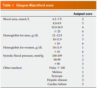
2) Glasgow-Blatchford score (GBS) ; 수혈의 필요성 예측률 높음
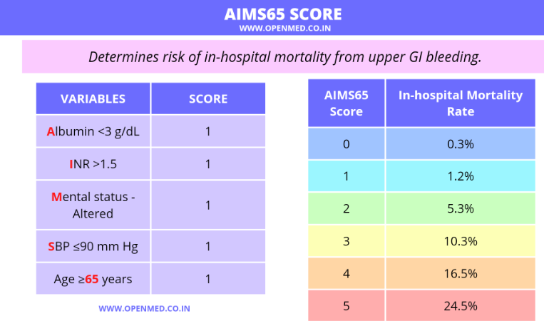
3) AIMS65 점수 ; 사망률 관련 높은 예측률
* The collected data were used for calculating the RS, GBS, and AIMS65 scores. The post-endoscopic clinical RS which is the most commonly used scoring system including age, signs of shock, comorbidities, endoscopic diagnosis, and evidence of bleeding was investigated in this study. The GBS includes the following parameters: blood urea nitrogen, hemoglobin, systolic blood pressure, pulse rate, presence of melena or syncope, hepatic disease, and heart failure. It can be calculated simply, as it does not require endoscopic findings or the determination of the degree of systemic disease. The AIMS65 score is a more simplified scoring system and it consists of five risk factors: hypoalbuminemia, age > 65 years, low systolic blood pressure, altered mental status, and prolonged prothrombin time.

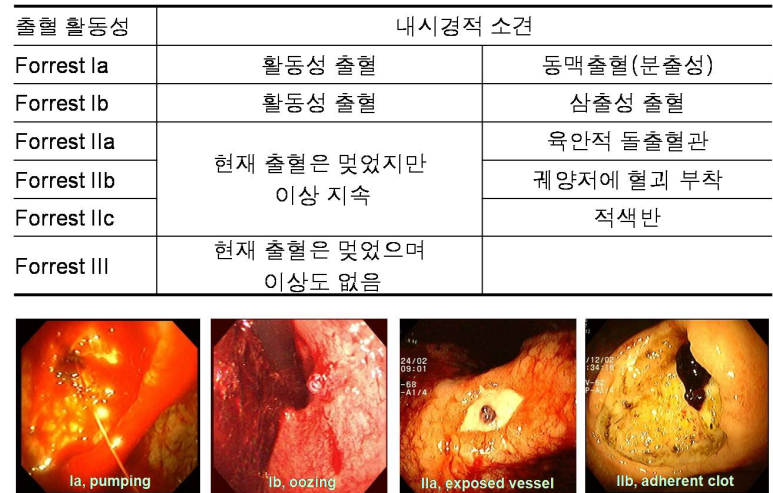
-재출혈에 연관된 인자 : Forrest Ia, Ib, IIa, IIb, NSAID 사용, co-morbidity 중 심혈관 질환
* Peptic ulcer bleeding was stratified according to endoscopic appearance using the Forrest classification (Ia, spurting bleeding; Ib, oozing bleeding; IIa, nonbleeding visible vessel; IIb, adherent clot; IIc, hematin on ulcer base; III, clean ulcer base)
endotoday
-Marginal ulcer : 위 부분 절제술 이후 관찰되는 위출혈
-Dieulafoy's lesion : 소화성 궤양에 비해 재출혈률이 유의하게 높음
EndoTODAY by Jun Heang Lee
[십이지장궤양이 아닙니다 (4). Dieulafoy lesion] Dieulafoy lesion은 위장관 어디에서나 발생할 수 있습니다. [Dieulafoy lesion에 의한 사망. 저절로 출혈하여 사망한 예와 검진 내시경 조직검사 후 사망한 예
endotoday.com
The characteristic endoscopic appearance of the Dieulafoy lesion is blood spurting or oozing from a pinpoint mucosal defect or, in the absence of bleeding, a clot without an associated surrounding ulcer. Occasionally, one can see a small 1- to 2-mm arterial vessel or a fibrin clot protruding from a normal looking mucosa. The mucosal defect typically occurs within an area of otherwise normal mucosa. There is no surrounding inflammation, exudates, or ecchymosis. Frequently, isolated intraluminal blood is suggestive of recent bleeding.
-refeeding period after endoscopic hemostasis : 내시경 지혈술 이후, 식이 24시간 이후 재개군과 48시간 이후 재개군으로 나누어 분석하였을때 30일 이내 재발성 출혈률은 24시간 이후 재개군에서 유의하게 높음